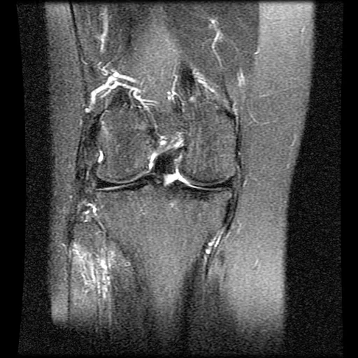
Explore the structures around my right knee, what the later reports described, and how a force through the joint could affect its outer side and the nerve that helps lift the foot.
A model for understanding. The bone and muscle surfaces come from a generic anatomical reference. The ligaments, tendons, and nerve are illustrative additions, not structures segmented from my magnetic resonance images. The records describe damage, but cannot establish the exact position, force, or sequence of the fall. Movements below are illustrative possibilities, not a reconstruction of what certainly happened.
RIGHT KNEE · FRONT / OUTER SIDEREFERENCE ANATOMY Illustrative soft tissue
Loading anatomical surfaces…
Reported injury · location approximate
Drag to orbit · Scroll or pinch to zoom · Arrow keys to rotate
Lower bone opacity to see the crossing ligaments inside the joint.
A backward force on the upper shin with the knee bent can load the posterior cruciate ligament. This alone does not explain every injury in the record.
02 / Explore a possible mechanism
What could have loaded these structures?
The recorded event is a trip while running, a fall onto the right knee, and immediate foot drop. The exact knee angle and foot position are unknown. Choose a possible loading direction to see the anatomical relationships; these are not measured simulations.
How to read the movement
Slider position is animation progress, not force, injury severity, or a safe range of movement. Coloured injury markers stay visible: moving back to the resting illustration does not mean the injuries have healed.
Use the outer-side view to see backward movement of the shin, and the front view to see the outer joint open. The visual combines later reported damage with hypothetical motion to explain relationships, not the timing of each injury.
03 / Separate the evidence from the illustration
What we know. What we cannot replay.
The documented pattern
The January summaries describe partial injuries to both cruciate ligaments, a detached biceps femoris insertion, a partial popliteus insertion injury, and marked nerve changes. The 31 January summary also describes a ruptured lateral collateral ligament and tissue compressing the nerve.
These structures sit close together, especially around the back and outer side of the knee. The model shows their relationship. It does not establish that they were injured in a particular order.
Why the foot was affected
The common peroneal nerve carries signals to muscles that lift the foot and toes. Its exposed route around the fibula makes it vulnerable to trauma. Stretch, direct impact, and compression are possible mechanisms. Immediate foot drop was recorded; later swelling or a neuroma should not be presented as if it had already formed at the moment of impact.
What remains unknown
The available material cannot determine the exact force, twist direction, whether the foot was planted, or whether there was a transient dislocation. The model does not assert a dislocation. It also does not show today's healing, surgical grafts, or a prognosis.
Bone and two muscle surfaces are drawn from BodyParts3D reference anatomy. Soft-tissue attachment points, tear markers, and movement are approximate. Menisci, cartilage, vessels, and most muscles are omitted; exposed bone surfaces do not mean cartilage was absent or injured. The model is not a biomechanical simulation. A surgeon could help assess which mechanism best fits the full original records.
Keep the images beside the explanation.
This model uses the report summaries already transcribed in this site. The rendered scan slices provide visual context; they were not used to diagnose additional injuries or recover patient-specific geometry.